Hearing and hearing aids
How does hearing work?
Hearing is a multi-layered process and therefore cannot be described by one scientific discipline alone.
First of all, physics and physical acoustics deal with the process of sound transmission and the associated hearing. The deeper the sound travels from the auricle into the interior of the ear and thus makes its way towards the brain, the more scientific fields come into play: anatomy, pathology, neurology, sensory physiology, psychoacoustics and psychology, to name just a few.
Georg von Békésy (1899 to 1972) made groundbreaking discoveries in the understanding of hearing. He received the Nobel Prize in Medicine for his research in 1962.

The outer ear
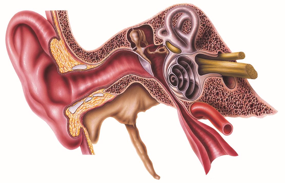
The auricle (pinna) picks up airborne sound from the environment. Its squiggly shape helps to analyze the direction of sound in a first step. This was already discovered by students in the 1960s, who filled their auricles with soft wax, rendering the shape of the auricles ineffective.
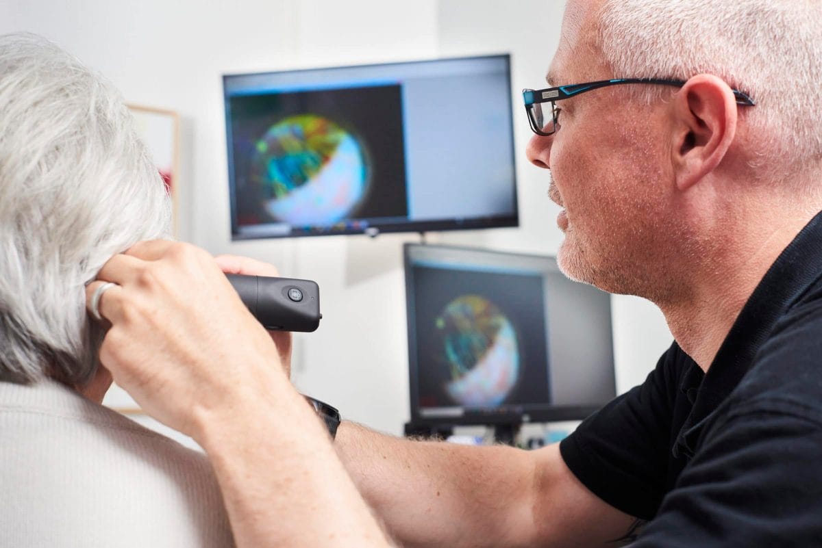
The sound collected by the auricle is conducted through the external auditory canal (meatus) to the inner ear. The ear canal is S-shaped, so that the direct free view from the outside to the eardrum is blocked without technical means. With the help of an otoscope, hearing care professionals can inspect this part of the ear. The visual impression of the eardrum alone provides important information that can influence the choice of the right hearing system. The ear canal ends after about 2.5 cm at the eardrum (Membrana tympani). An intact eardrum also seals the middle and inner ear behind it from penetrating air, water and pollution. In the outer ear are glands that produce earwax (cerumen). This has many tasks. It keeps the eardrum supple. It carries dirt out of the ear canal and also prevents infections in the ear through its disinfecting effect. Excessive production of cerumen can close the ear canal and must then be removed by an ENT specialist.
The middle ear
The eardrum marks the boundary between the outer ear and the middle ear. It's set into vibration by the sound waves in the auditory canal and transmits these vibrations to three ossicles in the middle ear: the malleus, incus and stapes. From the eardrum, sound is transmitted solely through this ossicular chain (ossicula auditus). The middle ear ends at the stapes footplate, which abuts the oval window of the cochlea.
To enable the ossicles to vibrate without loss, they are suspended by small muscles and ligaments inside the tympanic cavity (cavum typani). When exposed to strong sound, these muscles can block the oscillation of the ossicles and thus protect the ear from damage. The ossicular chain can be thought of as a mechanical transmission that amplifies sound transport. Additional sound amplification occurs because the area of the eardrum is about 14 times that of the oval window. Normally, sound from the air is reflected by water at 100 %. This middle ear design ensures that 60 % of the incoming sound reaches the fluid-filled inner ear. Technically, one therefore speaks of an impedance amplifier.
The inner ear
The inner ear begins at the oval window. While in hearing acoustics one speaks of sound conduction through the outer and middle ear up to this point, the inner ear is responsible for the so-called sound perception. From here, the sound waves are converted into electrical signals that are transmitted to the brain via the auditory nerve. The central organ of perception is the cochlea. Sound travels through the fluid-filled interior of the cochlea. If you unroll the 2.5 turns of the cochlea, you get a 3 cm long tube, whose cross-section is separated lengthwise into two chambers by the so-called basilar membrane. The basilar membrane is covered by the so-called organ of Corti. This is the actual stimulus-receiving hearing organ. According to hydrodynamic theory, the wave sets off in the direction of the tip of the snail, with the deflection and frequency increasing continuously. This wave is absorbed at the location of the highest amplitude of vibration. The frequency of the tone heard determines the path length to the absorption frequency: the higher the tone, the shorter the path. Each tone is thus assigned its own absorption site by its pitch, high tones in lower, lower tones in higher cochlear coils. At the different locations of the organ of Corti, nerve cells pick up the electrical signals from the sensory cilia, which are moved by the vibrating lymph fluid. Figuratively, one can imagine this as a cornfield through which the wind blows.
The inner hair cells are considered the actual sensory cells in the inner ear. They respond to this mechanical stimulus with action potentials on the conducting fibers of the auditory nerve. This leads to the auditory cortex, the control center of hearing in the cerebrum.
We have seen that the otoscopic (optical) analysis of a hearing care professional only reaches as far as the eardrum (outer ear). Through a professionally performed audiometry, however, our hearing care professionals can also draw conclusions about the function of the middle and inner ear. A hearing test, which we perform free of charge, is therefore worthwhile for you in several ways. It not only describes your hearing in general, but can also determine which part of the ear is not functioning properly. Let us make you an appointment for a hearing test today. Hearing test give

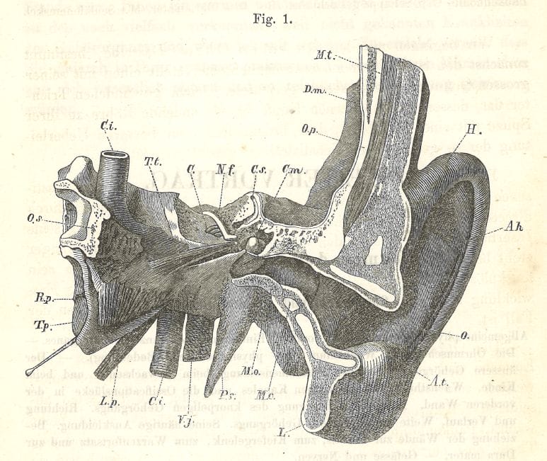
In May 1862, the textbook of ear medicine by Dr. Anton von Tröltsch was published.
historical retrospection
It is interesting to read that already at Tröltsch the present difference between sound-conducting part and sound-sensing part was known:
"The anatomist calls the sound-sensing apparatus, namely the spread of the auditory nerve in the labyrinth and the bone section containing these parts, the "inner ear"; in the other, more peripherally located apparatus, he distinguishes two divisions, the middle and the outer ear."
H - Helix (ear strip)
Ah - Anthelix (counter bar)
A.t. - Antitragus
L. - Lobulus auriculae (earlobe)
O. - outer ear opening and beginning of the auditory canal
M.c. - cut through cartilaginous ear canal
M.o. - cut through bony ear canal
P.s. - styloid process (elongated, pencil-shaped bone process)
V.j. - internal jugular vein ("internal jugular vein")
C.i. - internal carotid artery (internal carotid artery)
L.p. - levator palati (part of the palatal muscles)
T.p. - Tensor palati ("soft palate tensioner")
R.p. - Recesus pharyngis ("Rosenmüller space")
O.s. - cut through sphenoid bone
T.t. - Musculus tensor tympani ("muscle of the eardrum")
C. - Hearing cochlea (partially open)
N.f. - Facial nerve
C.s. - Upper semicircular canal
C.m. - Hammer head
O.p. - Scale of the temporal bone
D.m. - Dura mater (hard, outer meninges)
M.t. - temporal muscle

